


 183 / 352

179
MICROSTRUCTURAL ANALYSIS OF CRACK TIP REGION
Katarina Gerić
Faculty of Technical Sciences, Novi Sad, Serbia
gerick@uns.ns.ac.yu1. INTRODUCTION
The fracture develops from the crack tip. The investigation of crack propagation, the
analysis of the stress fields ahead the crack tip, and the microstructural analysis, including
fracture surface are inevitable parts in understand fracture process.
2. BRITTLE FRACTURE
The fracture is usually initiating from crack already present in material and developed
by crack growth. Local stress increase, caused by flaws in the material, significantly con-
tributes to fracture development. When the development of deformation is restrained the
fracture of metals is of brittle type in the Griffith sense. Anyhow, metal does not behave
in an ideally brittle manner and the plastically deformed zone on the crack tip occurs.
When its size is neglectible compared to component dimension and crack size, resulting
fracture can be considered as a brittle one. It is to underline that once started brittle
fracture will developed by cleavage mechanism, with no additional energy consumed.
Cleavage involves breaking bonds: the local stress must be sufficient to overcome the
cohesive strength of the material, which is higher than the remote stress required to cause
fracture. In order to initiate cleavage, there must be a local discontinuity ahead of the
macroscopic crack, sufficient to exceed the bond strength. Sufficiently sharp micro crack
can produce local stress concentration. Cottrell suggested that micro cracks by dislocation
interaction form at intersecting slip planes. However, more common mechanism for
micro crack formation in steel is connected with inclusions and second–phase particles.
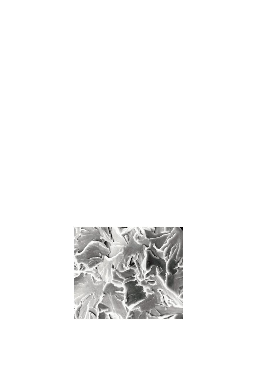
Figure 1 shows scanning electron microscope (SEM) fractograph of a cleavage fractu-
re in high-strength low-alloy (HSLA) steel /1/.
Figure 1: SEM fractograph of cleavage in steel (2000x)